

Predicting Alzheimer’s Progression from a Single Brain Scan: A New Milestone in AI-driven Diagnosis and Prognosis
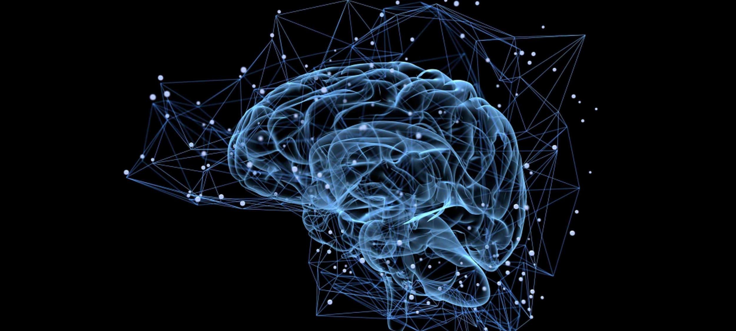
A study published in Nature Aging (May 18, 2026) reports a promising advance in Alzheimer’s disease (AD) diagnosis: an AI method that can predict not just whether someone has Alzheimer’s, but how their thinking and memory are likely to change over the next few years — all from one standard MRI brain scan and basic demographic information.
Why This Matters
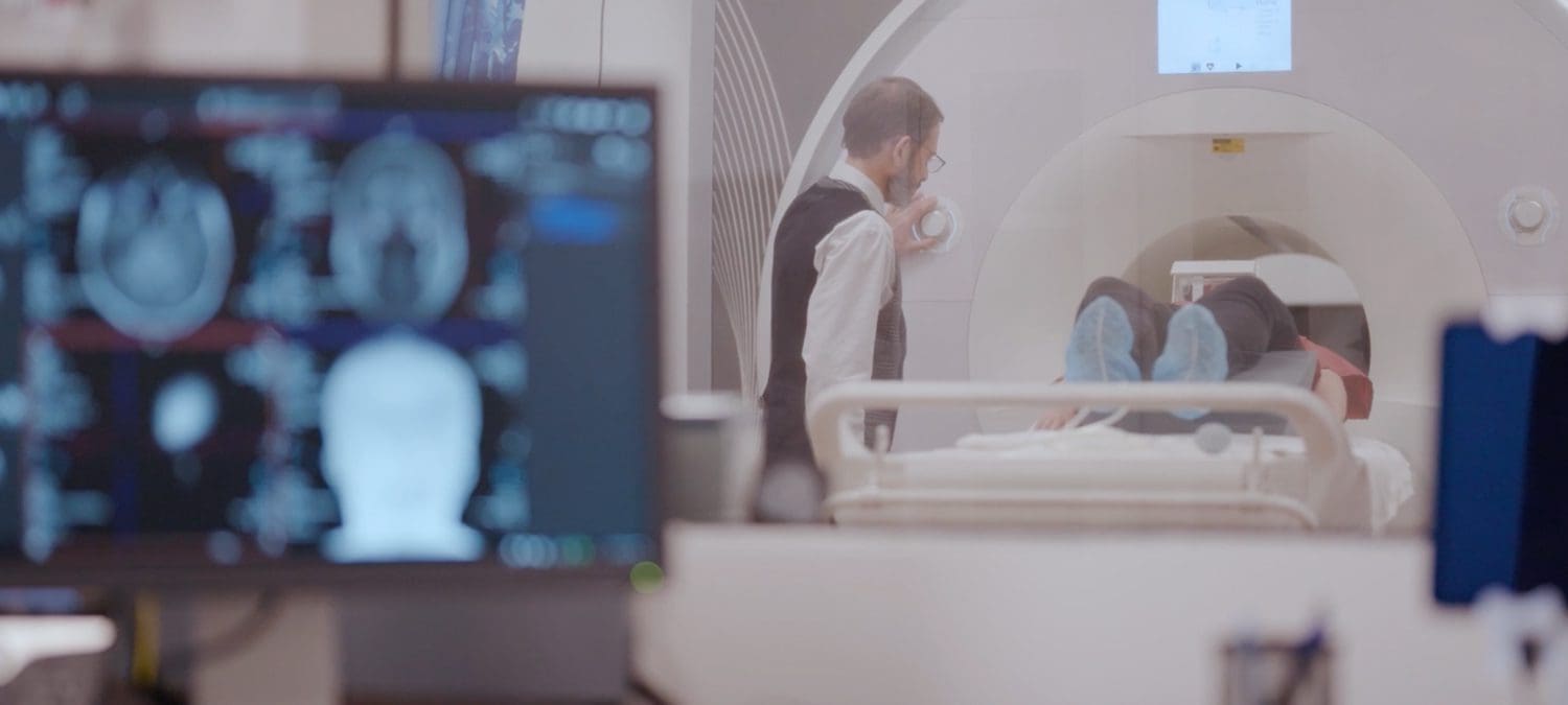
Alzheimer’s affects tens of millions worldwide. Doctors and researchers rely heavily on cognitive tests (like the ADAS-Cog) to assess severity and track progression. However, these tests are time-consuming, require specialized administration, and don’t always predict future decline accurately on their own. While brain imaging, especially MRI, is already standard in clinics, AI models have historically struggled to turn a single baseline scan into reliable predictions of continuous cognitive outcomes over time.
Previous efforts often needed expensive combinations of PET scans, multiple visits over time, blood tests, genetics, or labor-intensive manual image processing. This new approach aims for real-world usefulness by leveraging data that is available in ordinary clinical settings.
What the Researchers Did
The UCSF team developed a multitask deep learning framework that simultaneously performs multiple tasks from a single baseline MRI scan combined with basic demographic information (such as age, marital status, and education). The AI tool:
- Performs brain tissue segmentation (gray matter, white matter, CSF) to capture atrophy patterns.
- Predicts diagnostic category (AD vs. normal aging/MCI).
- Estimates current cognitive scores (ADAS-Cog).
- Forecasts future cognitive scores up to 36 months ahead.
To achieve this, they combined two powerful strategies: transfer learning (borrowing visual expertise from massive, pretrained medical imaging AI models) and domain-knowledge-driven customization (using a specialized architecture called a 3D U-Net). The U-Net helps ensure the AI’s predictions are guided by actual biological tissue changes rather than computational artifacts that can plague other AI methods.
This hybrid, knowledge-informed approach significantly outperformed standard AI benchmarks, achieving strong but not perfect predictive performance (for example, explaining roughly 80% of the variance in cognitive scores). The results demonstrated robust accuracy that held up even when tested on external validation datasets from entirely different patient populations.
Potential Benefits
- Earlier, personalized prognosis: One routine MRI could help clinicians estimate how quickly an individual patient might decline, allowing families more time to plan and access support.
- Accelerated clinical trials: By accurately identifying “rapid progressors” early on, researchers can design smaller, faster, and more cost-effective clinical trials for new therapeutics.
- Equitable access: Relies on widely available standard MRI, potentially reducing reliance on expensive/invasive tests.
- Scientific insight: Strengthens the link between structural changes and cognitive function
Important Caveats
- This is a significant research advance, but it is not yet a ready-to-use clinical tool. It will require validation across larger, more diverse patient populations, regulatory approval, and future integration with fluid biomarkers (like blood tests or cerebrospinal fluid markers for amyloid and tau) before it can be widely used for reliable diagnoses.
- MRI alone has limits. It captures structure but not the full pathology (amyloid, tau, inflammation, vascular factors). A multifaceted problem requires a multifaceted solution.
- While performance was strong in this study, real-world predictive accuracy can vary across different hospital systems, scanner manufacturers, and imaging protocols.
The Bottom Line
By transforming a single standard brain scan into a 36-month cognitive forecast, this study demonstrates the growing power of translation-focused AI to bridge the gap between complex neuroimaging data and meaningful, proactive patient care.
Expert Perspective
Jeffrey Luci, Ph.D., Research Assistant Professor and Technical Director of the Center for Advanced Human Brain Imaging Research (CAHBIR) within the Rutgers Brain Health Institute (BHI), commented, “The speed at which biomedical imaging methods are developed, while impressive using only legacy techniques, has rapidly accelerated with the addition of AI assistance. This work by colleagues at UCSF demonstrates one reason for that.”

“Brain segmentation has been a routine tool used for decades in neuroimaging applications, aiding in brain mapping and diagnosis & grading of disease. But without the insight provided by these models, it is not clear that these latent connections to other available data would have ever been discovered. Their mathematical analyses have greatly increased the value and utility of existing diagnostic tools and someday soon will result in real-world benefit to people who suffer from AD and other pathologies,” said Dr. Luci.
Reference
- Predicting categorical and continuous Alzheimer’s disease outcomes from a single MRI scan | Nature Aging
- https://brainhealthinstitute.rutgers.edu/our-centers/cahbir/
- https://sites.rutgers.edu/cahbir/
